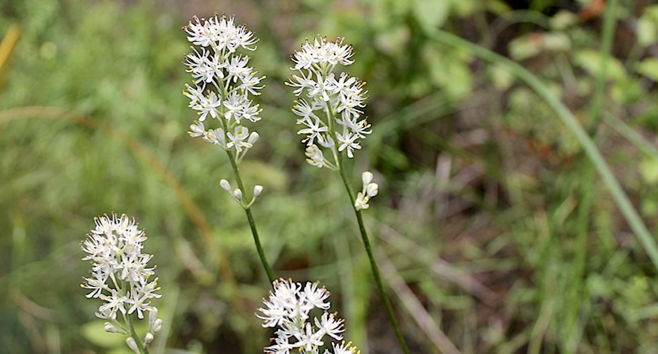
Iron-Air Batteries Powered by Rust Could Revolutionize Energy Storage By Using Only Iron, Water, and Air

Iron-air batteries for stable power – Credit: Form Energy

Edited with permission of EarthTalk® and E – The Environmental Magazine, Dear EarthTalk: What’s new regarding more efficient batteries that can help usher in a new age of renewable energy?

Batteries are everywhere—in your phone, your car—even the artificial organs many depend on for life. Fortunately, new innovations have increased the efficiency and sustainability of our ubiquitous batteries.

One of the most novel innovations unveiled recently is the iron-air battery system which usees rust to produce energy in a sustainable way.

The iron-air system from Form Energy is built from safe, low-cost, abundant materials—iron, water, and air—and uses no heavy or rare-earth metals. The company touts that approximately 80% of its components are sourced domestically from within the United States.

As air passes through the cathode (the negatively-charged portion of the battery) and reacts with the liquid, a water-based electrolyte, ions subsequently latch onto the positively-charged iron anode, producing rust. The movement of ions through this rust produces electricity, a process that can be repeated by continually un-rusting the battery after each reaction.

Form energy co-founder and Chief Scientist Yet-Ming Chiang notes the economic viability of iron-air batteries for large-scale usage: “Air is still free and iron is one of the most widely produced, lowest cost materials in the world.”

In Minnesota, a 1.5 megawatt pilot project was shown to be able to power 400 homes for 100 hours. It also successfully completed UL9540A safety testing, demonstrating the highest safety standards with no fire or thermal threats across all scenarios.

Besides iron-air batteries, solid-state batteries are what George Crabtree, director of the Joint Center for Energy Storage Research, believes to be “very likely… the next big thing at the commercial level.”

Solid-state batteries use electrolytes like argyrodite, garnet and perovskite that are more efficient than liquid-electrolytes in nearly all aspects: they’re lighter, take up less space and can hold more energy per unit of mass. These qualities make them effective for electrical vehicles and grid-scale energy storage.

However, researchers like University of Houston professor Yan Yao, who recently developed a glass-like electrolyte, are still looking for materials that fulfill all four factors for viability in the market: low-cost, easy-to-build, having a high degree of mechanical stability, and chemical stability.

With lithium-based batteries being so ubiquitous, some scientists are looking to improve on the existing model rather than supplanting it entirely. Batteries made out of lithium-sulfur, for example, exhibit four times greater energy density than traditional lithium batteries due to their usage of light, active materials.

Ultimately, innovations in batteries are a cornerstone to shaping a more sustainable future, making renewable energy more reliable and energy grids more stable.

EarthTalk® is produced by Roddy Scheer & Doug Moss for the 501(c)3 nonprofit EarthTalk. See more at emagazine.com. To donate, visit Earthtalk.org. Send questions to: question@earthtalk.org.
Read More........

Rapid BP fluctuations may signal risk of brain degeneration in elderly

(Photo: AI generated image/IANS)

New Delhi, (IANS) Older adults whose blood pressure fluctuates widely may indicate a greater risk of brain shrinkage and nerve cell injury, according to a new study.

The study, published in the Journal of Alzheimer’s Disease, reveals that short-term “dynamic instability” in blood pressure -- moment-to-moment changes measured over just minutes -- is linked to loss of brain tissue in regions critical for memory and cognition, as well as to blood biomarkers of nerve cell damage.

“Our findings show that even when average blood pressure is normal, instability from one heartbeat to the next may place stress on the brain,” said Daniel Nation, from the University of Southern California’s Leonard Davis School of Gerontology.

“These moment-to-moment swings appear to be associated with the same kinds of brain changes we see in early neurodegeneration,” added the Professor of Gerontology and Medicine.

While high average blood pressure has long been known to increase the risk of dementia, this study focuses on blood pressure variability, or how much blood pressure rises and falls over short time periods.

Recent evidence suggests that such fluctuations can strain small blood vessels in the brain and reduce their ability to deliver steady blood flow.

In this study, the researchers combined two complementary measures: Average Real Variability (ARV) and Arterial Stiffness Index (ASI).

ARV captures how much systolic blood pressure (the top number in a blood pressure reading) changes between each heartbeat, while ASI reflects how flexible or stiff the arteries are as they respond to those changes in pressure.

Together, these measures indicate how much blood flow changes over a short period of time, or what the researchers call “blood pressure dynamic instability.”

"This study suggests that excessive fluctuations could be a sign of vascular ageing that contributes to brain injury,” Nation explained.

The team conducted MRI scans of 105 community-dwelling older adults between the ages of 55 and 89 who were generally healthy and had no major neurological disease.

Participants with both high ARV and high ASI were found to have smaller hippocampal and entorhinal cortex volumes -- brain regions vital for learning and memory, also the first affected by Alzheimer’s disease.

Blood samples showed that the same individuals also had higher levels of neurofilament light (NfL), a blood-based marker that rises when nerve cells are damaged.The findings open a new window into how cardiovascular changes contribute to cognitive decline and may offer novel prevention strategies. Rapid BP fluctuations may signal risk of brain degeneration in elderly | MorungExpress | morungexpress.com
Read More........

First Antidote for Carbon Monoxide Poisoning 'Cleans' Blood in Minutes

A plug-in carbon monoxide detector – credit Fastily CC 4.0. BY-SA

It’s invisible, it’s lethal, and it’s been with us for decades, but carbon monoxide poisoning can now be fought with the first-ever antidote that rapidly removes the toxic molecule from the bloodstream.

Carbon monoxide or CO, poisoning accounts for 50,000 emergency room visits in the US each year and causes about 1,500 deaths, each one being lamented as a shameful waste and tragic oversight.

Currently, the only treatments for CO poisoning are oxygen-based therapies, which help the body eliminate the toxic gas. However, even with treatment, nearly half of survivors suffer long-term heart and brain damage. This has created an urgent need for faster, more effective interventions.

In a study published by University of Maryland School of Medicine (UMSOM) in PNAS, the research team developed a new engineered protein-based therapy called RcoM-HBD-CCC, which acts like a sponge to soak up carbon monoxide from the blood.

RcoM (short for “regulator of metabolism”) is a natural protein isolated from the bacterium Paraburkholderia xenovorans which uses it to sense minute levels of carbon monoxide in its environment.

The researchers engineered a version that is highly selective, grabbing CO without interfering with oxygen or other important molecules in the bloodstream like nitric oxide, which is vital for the regulation of blood pressure.

In tests on mice, the new therapy worked quickly to remove CO from red blood cells and was safely flushed out of the body through urine.

CO is known commonly as the “silent killer,” because this odorless, invisible gas, typically released from combustion sources, including stovetops, propane heaters, car exhausts, and firewood, poisons in a gradual manner that isn’t immediately obvious to the victim.

In healthy bodies, oxygen inhaled from the air binds to the protein hemoglobin on the surface of red blood cells, which then ferry the oxygen to all the tissues of the body. CO however, competes with oxygen for hemoglobin. It enters the bloodstream and binds to hemoglobin with a 200 to 400-fold greater affinity than oxygen. That means CO hogs most of the hemoglobin seats, so not enough oxygen molecules can get a ride to the tissues that need them.

Currently, the only available treatments for carbon monoxide poisoning involve giving 100% pure oxygen, sometimes under pressure in a hyperbaric chamber.

All too often, patients are not transported, diagnosed, and treated in time to reverse the effects of CO poisoning, which can cause lasting cardiac and neurological injuries or even death.

Infused in the bloodstream, scavenger hemoproteins like RcoM-HBD-CCC rapidly bind to carbon monoxide molecules, reducing the time it takes to clear half of the carbon monoxide in the blood to less than a minute, compared to more than hour with pure oxygen therapy and five hours without any treatment.

A potential drawback the researchers were aware of is that so-called “scavengers” like RcoM also have an affinity for oxygen, and so may uptake the nitric oxide mentioned earlier. This can cause wild and potentially unsafe changes in blood pressure, but RcoM-HBD-CCC caused no such side effects.

“Unlike other protein-based treatments, we found the compound caused only minimal changes in blood pressure, which was an exciting finding and raised the potential for this new molecule to have clinical applications,” said study corresponding author Mark T. Gladwin, MD, Dean of UMSOM.

“This has the potential to become a rapid, intravenous antidote for carbon monoxide that could be given in the emergency department or even in the field by first-responders.”Future studies will likely include more pre-clinical research to determine the safe and effective dosage range for RcoM-HBD-CCC in treating carbon monoxide poisoning. It could also form the basis for new research in other fields, including as an oxygen delivery therapy or blood substitute. This could include hemorrhagic shock, acute respiratory distress syndrome (ARDS), severe anemias, and the preservation of organs for transplantation. First Antidote for Carbon Monoxide Poisoning 'Cleans' Blood in Minutes
Read More........

Six Baby Cheetahs Born in the Richmond Zoo's Prolific Breeding Program – 167 Cats Since 2013 (WATCH)

Six cheetah cubs with their mother –Courtesy of Metro Richmond Zoo

At a zoo in Richmond, a raucous litter of cheetah cubs is delighting onlookers having spent the summer months growing in secret.

One of the largest litters born at the zoo, the three-month-old cubs are hitting milestones and progressing well towards adolescence.

Named after African capitals, such as Lusaka, Kampala, and Cairo, the cubs were born in April but were kept hidden away to develop in peace through their most sensitive period with mother Zuri.


Sprinting, jumping on each other, and investigating every object in their enclosure, they seem well on their way to becoming the world’s fastest land animal.

“Some are shy; one is bold and brave — that’s Lusaka,” said Kristina Coonley, a lead zookeeper at the Metro Richmond Zoo, Virginia. “[Cairo] is always the last one out and the last one to come in.”

Though just the 99th most populated city in America, the Richmond Zoo nevertheless boasts one of the country’s most prolific and successful cheetah breeding program for purposes of conservation. Zuri’s litter of 6 takes the total number of captive-born cubs in the program’s history to 167 since 2013.

Coonley told the Washington Post that the Cheetah Conservation Center looks carefully and genetics and disposition when deciding which of their cats are suitable for breeding. Zuri is 5 years old, and has already mothered one litter. Her partner was Ramses, a 13-year-old sire of 25 other cubs.

Though losing out on conservation column inches to the likes of the lion and tiger, cheetah are considered Vulnerable by the IUCN. With around 6,500 individuals across the whole of Africa, a relic populations in Iran, and a diminishing introduced population in India, they are the most at-risk of the African big cats because of their need for space befitting the world’s greatest sprinters.It’s no bad thing then that the Metro Richmond Zoo has seen even bigger litters than these 6, with a resident female last year bringing 9 little mohawked babies into the world. Six Baby Cheetahs Born in the Richmond Zoo's Prolific Breeding Program – 167 Cats Since 2013 (WATCH)
Read More........

Researchers Blown Away After Finding Jaguar Population up 30% Across Mexico

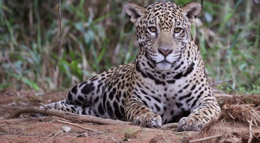
A jaguar on the Piquiri river – credit, Charles J. Sharp from Sharp Photography CC 4.0. BY-SA via Wikimedia

Imagine sitting in southern Texas and knowing that in less than a day you could drive to se the world’s third largest cat.

That is absolutely the reality in Mexico today, following a second national jaguar population census which found this charismatic cat is actually increasing in numbers across the country, reaching around 5,300 animals.

Gerardo Ceballos and colleagues conducted the first census in 2010, hoping to grasp more or less the gravity of the risk of extinction faced by the jaguar. They were estimating they’d find around 1,000 in the whole of Mexico.

But rather than the risk of extinction, their results conveyed a different narrative. They found four-times as many cats as they expected.

“It was a great surprise, terrific news,” Ceballos said. “Obviously 4,000 means the species is still in danger of extinction, but 4,000 is a lot better than 1,000.”

Then in 2025, Ceballos completed a second survey, employing over 50 national and local research institutions and community leaders, who together set up 920 motion-activated camera traps.

Staggered again, the count showed that in 15 years, jaguar numbers had increased 30%. It turns out that even though the twenty-teens saw hundreds of thousands of acres of forest cleared, there were almost as many jaguars in Mexico as there are cheetahs on the entire African continent.

“The fact that the country has managed to maintain and increase its population over the last 14 years is extraordinary,” Ceballos told the Guardian. “For me it’s great news for the country. Mexico and the world need good news.”

The populations came in as follows: the Yucatán peninsula region had the most (1,699), followed by the south Pacific area (1,541), north-east and central Mexico (813), the north Pacific (733) and the central Pacific coast (540).

The jaguar is a compact, muscular predator, with exceptionally strong jaws that allow it to pierce the shells of turtles and the hides of crocodilians. They’re good tree climbers, avid swimmers, and will even hunt in the water. Like tigers, it employs a stalk and ambush hunting strategy, and is considered peerless in terms of its catch rate.

In short, and by comparison, the jaguar is maybe the most versatile and adaptable of any of the big cats.

This gives the animal an advantage in the gradually shrinking forests of Mexico.Ceballos told the Guardian that the spotted hunter would face multiple threats, including from the continued construction of new highways across Mexico, as well as habitat loss and zoonic diseases being passed to them via livestock which they occasional poach. Researchers Blown Away After Finding Jaguar Population up 30% Across Mexico
Read More........

Scientists Find Answer to Sea Star Population Devastated by Pathogen Along the California Coast

A sunflower sea star – credit, Ed Bierman CC 2.0.

For years, a wasting disease has been turning sea stars to goo off the California coast. Scientists now finally know the cause, and are beginning to fight back.

Whether it has over 20 arms like the sunflower sea star, or just 5, billions of Pacific sea stars were being wiped out by an unknown assailant.

After four years of experiments from a huge collaborative effort led by the Hakai Institute, biologists finally identified the culprit: a kind of bacteria called Vibrio.


Devastating to coral, shellfish, and human beings, this strain of Vibrio has been labeled FHCF-3. The scientists determined it was the cause of the epidemic by examining what might be called the sea star’s blood. It doesn’t have blood as we would recognize it, but a circulatory fluid called coelomic fluid.

As to what is causing the spread of FHCF-3, ranging from Washington state down to the Baja Peninsula, the scientists point to warming waters.

“We have evidence that there is a link between increasing ocean temperatures and this sea star wasting disease epidemic,” said Melanie Prentice, one of the co-authors of the paper published on the discovery in Nature, to CBS News.

Sunflower sea stars, one of the species that’s been most affected, are voracious eaters of sea urchins. This slow motion game of lion and gazelle plays out on the seafloor and on reefs, and is a major cog in the overall machine of marine ecosystem stability.

Themselves voracious eaters of kelp, the urchins were unleashed following the sea star’s decline, and like the bacteria that decimated the sea stars, the urchins devastated the kelp.

With the cause identified, a large collaboration involving Prentice’s Hakai Institute, as well as the universities of British Columbia and Washington, the Nature Conservancy, Tula Foundation, US Geological Survey, and the Washington Department of Fish and Wildlife, are beginning to plan strategies for the sea stars’ recovery.

A breeding program for sunflower stars was set up between the Aquarium of the Pacific, the Birch Aquarium, the San Diego Zoo Wildlife Alliance, and the Sunflower Star Laboratory. Hundreds have already been raised, and biologists can now screen for the pathogen routinely.

Some of the juveniles are living in these aquariums, where members of the public can learn about the sea stars’ struggle to survive, and the critical role they play in the ecosystem.


WATCH the story below from CBS News’ ‘Project Earth’ segment…

Read More........

US scientists developing single-dose vaccines for HIV, Covid


New Delhi, August 28 (IANS) A team of scientists in the US is working to develop vaccines that can protect against HIV, Covid, and potentially other diseases, with a single dose.

The team from MIT and the Scripps Research Institute treated mice with a vaccine that combines two different adjuvants, materials that help stimulate the immune system. They found that the combination helped generate more robust immune responses.

The dual-adjuvant vaccine was found to accumulate in the lymph nodes, where white blood cells known as B cells encounter antigens and undergo rapid mutations that generate new antibodies.

The vaccine’s antigens remained there for up to a month -- allowing the immune system to build up a much greater number and diversity of antibodies against the HIV protein than the vaccine given alone or with one adjuvant.

According to MIT professor J. Christopher Love, the approach may mimic what occurs during a natural infection and could lead to an immune response so strong and broad that vaccines only need to be given once.

“It offers the opportunity to engineer new formulations for these types of vaccines across a wide range of different diseases, such as influenza, SARS-CoV-2, or other pandemic outbreaks,” Love said.

Separately, Russia’s Covid-19 vaccine maker is also set to develop an mRNA-based HIV vaccine, RIA Novosti, the Russian Information Agency, said.

The vaccine to be developed by Gamaleya Center in Moscow, which will trigger an immune response against the AIDS causing virus, could be ready in around two years.

The much-anticipated HIV vaccine is based on mRNA technology, the head of the institute’s epidemiology department, Vladimir Gushchin, told RIA Novosti on Wednesday.

“We are currently at the stage of creating the first antigens that will generate a broadly neutralising immune response. The success of the vaccine will depend on whether the immunogen we use can trigger a response capable of protecting against all variants,” said Vladimir Gushchin, epidemiologist at the Centre.The Gamaleya Center also developed Sputnik V, one of the world’s first Covid-19 vaccines, which was rolled out in August 2020. The shot, showing up to 97.8 per cent efficacy with no serious side effects, has been approved in nearly 70 countries. US scientists developing single-dose vaccines for HIV, Covid | MorungExpress | morungexpress.com
Read More........

Why Do Animals Play? Because They Need To Play – Just Like Children Do

Photo by Tambako The Jaguar, CC license

As much as it’s a time for growing and learning, childhood is also a time for unabashed joy. Pastimes like careening down a snowy hillside on your sled, flying off a rope swing into a cool lake on a hot summer day, or even just a game of catch are part and parcel of growing up.

But the joys of playtime aren’t just reserved for human kids—animal offspring are just as likely to get into the act as well, and some of their activities are startlingly similar to our own.

Young ravens hold body-surfing “competitions” down the slopes of wintery rooftops; juvenile elephants create impromptu waterslides along muddy riverbanks; herring gulls engage in their own version of airborne hacky sack substituting seashells for bean-filled projectiles.

Scientists believe that for certain animal species, some fun and games is strictly that—play for the sake of play—but as with humans, other forms of diversion are preparing youngsters for the rigors of adulthood.

“Play is essential to development because it contributes to the cognitive, physical, social, and emotional well-being of children and youth,” wrote Kenneth R. Ginsburg in the American Journal of Pediatrics. “Play also offers an ideal opportunity for parents to engage fully with their children.”

Those same tenets, it seems, hold true in the animal kingdom as well.

“Horses…are known to engage in play almost as soon as they are born. Once they can walk, they immediately start to gallop, frolic and buck, again, honing the motor skills they may need when they’re mature,” notes BBC Earth.
Play with purpose

But along with social and motor skills, play also teaches animals essential hunting and survival skills.

Inge Wallmrod

While the antics of cute cavorting kittens is the stuff that’s spawned a myriad of viral videos, whether it’s an opportunity to take down an errant mouse or to avoid harm in the face of unexpected danger, their ninja-like antics may in fact be helping kittens learn to be ready when life hands them a surprise.

Even natural-born predators, such as kestrels, use play to hone their hunting skills by practicing with targets that look like real prey when they’re young.

In the oceans, dolphins chase underwater air rings to fine-tune their sonar skills.

And while it’s unclear why bear cubs are so quintessentially playful, zoologists believe at least some of their shenanigans have a more serious purpose that aids in their survival as adults.

One of the most important teaching aspects of play is socialization. These days, for human kids, that usually means the basics like learning to share, teamwork, and knowing boundaries.

For animals, especially those that live in packs, flocks, or herds, play (often in the form of play fighting) imparts an understanding of where each animal fits into the community hierarchy.

In ways that are remarkably similar to the training children of traditional tribal cultures receive, it is through the rules of play that lion cubs, kangaroo joeys, and wolf pups discover and establish the roles they’ll be expected to perform as adults.

But for animals, not all socializing play is about fighting or establishing dominance. Some of it’s about learning to be better parents—and that involves playing with dolls. While they might lack a perambulator and a fancy wardrobe, female chimpanzees are known to lavish their doll babies with love and emulate their own mothers’ attentive care.

So whether it’s frolicking in the pasture, hanging from a tree, or rollicking in the surf, it seems that play will always be an intrinsic—and fun—part of both human and animal development.And we’re pretty sure when those ninja-kitten TikTok stars stop climbing that curtain, they’ll be thrilled to hear about it. Why Do Animals Play? Because They Need To Play – Just Like Children Do:
Read More........

Retooled Cancer Drugs Eliminated Aggressive Tumors in ‘Remarkable’ New Trial


Cancer drugs that have been used for two decades were retooled until they were able to eliminate aggressive tumors in a “remarkable” clinical trial.

Two of the patients—one with the deadliest form of skin cancer called melanoma and another with breast cancer—were told their tumors disappeared completely.

Scientists at Rockefeller University in New York engineered an upgrade to an antibody that improved a class of drugs—called CD40 agonist antibodies—which have struggled to make good on their early promise, but showed great potential.

While effectively activating the immune system to kill cancer cells in animal models, the drugs had only “limited” impact on humans, while also triggering dangerous adverse reactions.

So, five years ago, the team at the New York university engineered an enhanced CD40 agonist antibody so that it improved its efficiency and limited any serious side effects for mice, with the next step being a clinical trial with cancer patients.

The results from the phase 1 clinical trial of the drug, dubbed 2141-V11, showed that six out of 12 cancer patients saw their tumors shrink, including two that saw them disappear completely.

“Seeing these significant shrinkages and even complete remission in such a small subset of patients is quite remarkable,” said study first author Dr. Juan Osorio.

He said the effect wasn’t limited to tumors that were injected with the drug; tumors elsewhere in the body either got smaller or were destroyed by immune cells.

“This effect—where you inject locally but see a systemic response—that’s not something seen very often in any clinical treatment,” said Professor Jeffrey Ravetch who oversaw the study.

“It’s another very dramatic and unexpected result from our trial.”

Oral squamous cancer cell (white) being attacked by two cytotoxic T cells (red) – Credit: NIH

He explained that CD40 is a cell surface receptor and member of the tumor necrosis factor (TNF) receptor “superfamily”—proteins that are largely expressed by immune cells. When triggered, CD40 prompts the rest of immune system to spring into action, promoting anti-tumor immunity and developing tumor-specific T cell responses.

In 2018, Prof. Ravetch’s lab engineered the 2141-V11, a CD40 antibody that binds tightly to human CD40 receptors and is modified to enhance its cross-linking by also engaging a specific Fc receptor.

It proved to be 10 times more powerful in its capacity to elicit an anti-tumor immune response.

The research team then changed how they administered the drug. When previously given intravenously, too many non-cancerous cells picked it up, leading to the well-known toxic side effects.

They instead injected the drug directly into tumors. When they did that, they saw “only mild toxicity”, said Prof. Ravetch.

The new trial included 12 patients who had various types of cancer, and of those 12, six experienced systemic tumor reduction, of which two had their cancers (notorious for being aggressive and recurring) disappear entirely.

“The melanoma patient had dozens of metastatic tumors on her leg and foot, and we injected just one tumor up on her thigh. After multiple injections of that one tumor, all the other tumors disappeared,” Ravetch said.

“The same thing happened in the patient with metastatic breast cancer, who also had tumors in her skin, liver, and lung. And even though we only injected the skin tumor, we saw all the tumors disappear.”

Tissue samples from the tumor sites revealed the immune activity that the drug stimulated.

“We were quite surprised to see that the tumors became full of immune cells—including different types of dendritic cells, T cells, and mature B cells—that formed aggregates resembling something like a lymph node,” said Dr. Osorio.

“The drug creates an immune micro-environment within the tumor, and essentially replaces the tumor with these tertiary lymphoid structures, which are associated with improved prognosis and response to immunotherapy.”

The team also found TLS in the tumors they didn’t inject.

“Once the immune system identifies the cancer cells, immune cells migrate to the non-injected tumor sites,” said Dr. Osorio.

The findings, published in the journal Cancer Cell, sparked several other clinical trials that the Ravetch lab is currently working on with researchers at Memorial Sloan Kettering and Duke University.

The trials are investigating 2141-V11’s effect on specific cancers, including bladder cancer, prostate cancer, and glioblastoma—all aggressive and hard to treat.Nearly 200 people are enrolled in the various studies that the researchers hope will explain why some patients respond to 2141-V11 and others do not—and how to potentially change that.Retooled Cancer Drugs Eliminated Aggressive Tumors in ‘Remarkable’ New Trial
Read More........

AI can help detect early larynx cancer from sound of voice: Study


New Delhi, (IANS): A team of US scientists showed that Artificial Intelligence (AI) can help detect early larynx or voice box cancer from the sound of the patient’s voice.

Cancer of the voice box is an important public health burden. In 2021, there were an estimated 1.1 million cases of laryngeal cancer worldwide, and approximately 100,000 people died from it.

Risk factors include smoking, alcohol abuse, and infection with human papillomavirus.

The prognosis for laryngeal cancer ranges from 35 per cent to 78 per cent survival over five years when treated, depending on the tumour’s stage and its location within the voice box.

Now, researchers from the Oregon Health & Science University showed that abnormalities of the vocal folds can be detected from the sound of the voice using AI.

Such ‘vocal fold lesions’ can be benign, like nodules or polyps, but may also represent the early stages of laryngeal cancer.

These proof-of-principle results open the door for a new application of AI: namely, to recognise the early warning stages of laryngeal cancer from voice recordings, said the team in the paper published in the journal Frontiers in Digital Health.

“Here we show that with this dataset we could use vocal biomarkers to distinguish voices from patients with vocal fold lesions from those without such lesions,” said Dr Phillip Jenkins, postdoctoral fellow in clinical informatics at Oregon.

In the study, Jenkins and team analysed variations in tone, pitch, volume, and clarity with 12,523 voice recordings of 306 participants from across North America.

A minority were from patients with known laryngeal cancer, benign vocal fold lesions, or two other conditions of the voice box: spasmodic dysphonia and unilateral vocal fold paralysis.

The researchers focused on differences in a number of acoustic features of the voice: for example, the mean fundamental frequency (pitch); jitter, variation in pitch within speech; shimmer, variation of the amplitude; and the harmonic-to-noise ratio, a measure of the relation between harmonic and noise components of speech.

They found marked differences in the harmonic-to-noise ratio and fundamental frequency between men without any voice disorder, men with benign vocal fold lesions, and men with laryngeal cancer.

They didn’t find any informative acoustic features among women, but it is possible that a larger dataset would reveal such differences.Variation in the harmonic-to-noise ratio can be helpful to monitor the clinical evolution of vocal fold lesions, and to detect laryngeal cancer at an early stage, at least in men, the researchers said. AI can help detect early larynx cancer from sound of voice: Study | MorungExpress | morungexpress.com
Read More........

Scientists Define a Color Never Before Seen by Human Eyes, Called 'Olo'–a Blue-Green of Intense Saturation

Photo by Hamish on Unsplash

An experiment in human photoreceptors allowed scientists to recently define a new color, imperceptible by the human eye, that lies along the blue-green spectrum but is different from the two.

The team, who experimented on themselves and others, hope their findings could one day help improve tools for studying color blindness or lead to new technologies for creating colors in digital imagery.

“Theoretically, novel colors are possible through bypassing the constraints set by the cone spectral sensitivities…” the authors write in their abstract. “In practice, we confirm a partial expansion of colorspace toward that theoretical ideal.”

The team from University of California, Berkeley and the University of Washington used pioneering laser technology which they called “Oz” to “directly control the human eye’s photoreceptor activity via cell-by-cell light delivery.”

Color is generated in our vision through the transmission of light in cells called photoreceptors. Eye tissue contain a series of cones for this task, and the cones are labeled as L, S, or M cones.

In normal color vision, the authors explain, any light that stimulates an M cone cell must also stimulate its neighboring L and/or S cones because the M cone spectral response function lies between that of the L and S cones.

“However, Oz stimulation can by definition target light to only M cones and not L or S, which in principle would send a color signal to the brain that never occurs in natural vision,” they add.

Described as a kind of blue-green with “unprecedented saturation” the new color, which the researchers named “olo” was confirmed as being beyond the normal blue-green spectrum by each participant who saw it, as they needed to add substantial amounts of white for olo to fit somewhere within that spectrum.

“The Oz system represents a new experimental platform in vision science, aiming to control photo receptor activation with great precision,” the study says.


Although the authors are confidant that olo has never been seen before by humans, the spectrum of blue-green has received international attention before as a field of vision discovery.

A groundbreaking study of the Himba people in Namibia conducted in 2005 and published in journal of the American Psychological Association demonstrated that these traditional landowners seemed to perceive various colors as the same because they used the same word for them. A grouping of colors we in the West would separate into pink, red, and orange, is all serandu to them.

That was only half of the cause for fascination with the study. The other half came from the Himba people’s unbelievable sensitivity to the blue-green spectrum, such that they could reliably pick out the fainest differences in green that Western viewers by comparison missed.

This also corresponded with more words for shades of green which Westerners would never bother specifying, and in fact, the Himba had a harder time pointing out that a blue square was different from green squares when shown a chart, but could reliably select the square of a slightly different shade of green to the rest.But then it got even stranger. Further studies in the following years included genetic testing on the Himba, and it showed they possess an increased number of cone cells in their eyes. This higher density of cones enables them to perceive more shades and nuances of color than the average person, according to the lead author of the genetic research. Scientists Define a Color Never Before Seen by Human Eyes, Called 'Olo'–a Blue-Green of Intense Saturation
Read More........

Discovery of Genetically-Varied Worms in Chernobyl Could Help Human Cancer Research

Worms collected in the Chornobyl Exclusion Zone – SWNS / New York University

The 1986 disaster at the Chernobyl nuclear power plant transformed the surrounding area into the most radioactive landscape on Earth, and now the discovery of a worm that seems to be right at home in the rads is believed to be a boon for human cancer research.

Though humans were evacuated after the meltdown of Reactor 4, many plants and animals continued to live in the region, despite the high levels of radiation that have persisted to our time.

In recent years, researchers have found that some animals living in the Chernobyl Exclusion Zone are physically and genetically different from their counterparts elsewhere, raising questions about the impact of chronic radiation on DNA.

In particular, a new study led by researchers at New York University finds that exposure to chronic radiation from Chernobyl has not damaged the genomes of microscopic worms living there today, and the team suggests the invertebrates have become exceptionally resilient.

The finding could offer clues as to why humans with a genetic predisposition to cancer develop the disease, while others do not.

“Chernobyl was a tragedy of incomprehensible scale, but we still don’t have a great grasp on the effects of the disaster on local populations,” said Sophia Tintori, a postdoctoral associate in the Department of Biology at NYU and the first author of the study, published in the Proceedings of the National Academy of Sciences.

“Did the sudden environmental shift select for species, or even individuals within a species, that are naturally more resistant to ionizing radiation?”

Tintori and her colleagues turned to nematodes, tiny worms with simple genomes and rapid reproduction, which makes them particularly useful for understanding basic biological phenomena.

“These worms live everywhere, and they live quickly, so they go through dozens of generations of evolution while a typical vertebrate is still putting on its shoes,” said Matthew Rockman, a professor of biology at NYU and the study’s senior author.

“I had seen footage of the Exclusion Zone and was surprised by how lush and overgrown it looked—I’d never thought of it as teeming with life,” added Tintori. “If I want to find worms that are particularly tolerant to radiation exposure, this is a landscape that might have already selected for that.”

In collaboration with scientists in Ukraine and U.S. colleagues, including biologist Timothy Mousseau of the University of South Carolina, who studies the effects of radiation from the Chernobyl and Fukushima disasters, Tintori and Rockman visited the Chernobyl Exclusion Zone in 2019 to see if chronic radiation has had a detectable impact on the region’s worms.

With Geiger counters in hand to measure local levels of radiation and personal protective gear to guard against radioactive dust, they gathered worms from samples of soil, rotting fruit, and other organic material.
The ruins of Reactor 4, Chernobyl Exclusion Zone. credit Matt Shalvatis – CC BY-4.0. SA

Worms were collected from locations throughout the zone with different amounts of radiation, ranging from low levels on par with New York City (negligibly radioactive) to high-radiation sites on par with outer space (dangerous for humans, but of unclear if it would be dangerous to worms).

After collecting samples in the field, the team brought them to Mousseau’s field lab in a former residential home in Chernobyl, where they separated hundreds of nematodes from the soil or fruit. From there, they headed to a Kyiv hotel where, using travel microscopes, they isolated and established cultures from each worm.

Back in the lab at NYU, the researchers continued studying the worms by freezing them.

“We can cryopreserve worms, and then thaw them for study later. That means that we can stop evolution from happening in the lab, something impossible with most other animal models, and very valuable when we want to compare animals that have experienced different evolutionary histories,” said Rockman.

They focused their analyses on 15 worms of a nematode species called Oscheius tipulae, which has been used in genetic and evolutionary studies. They sequenced the genomes of the 15 O. tipulae worms from Chernobyl and compared them with the genomes of five O. tipulae from other parts of the world.

The researchers were surprised to find that, using several different analyses, they could not detect a signature of radiation damage on the genomes of the worms from Chernobyl.

“This doesn’t mean that Chernobyl is safe—it more likely means that nematodes are really resilient animals and can withstand extreme conditions,” noted Tintori. “We also don’t know how long each of the worms we collected was in the Zone, so we can’t be sure exactly what level of exposure each worm and its ancestors received over the past four decades.”

Wondering whether the lack of genetic signature was because the worms living in Chernobyl are unusually effective at protecting or repairing their DNA, the researchers designed a system to compare how quickly populations of worms grow and used it to measure how sensitive the descendants of each of the 20 genetically distinct worms were to different types of DNA damage.

The surprise in this story is that while the lineages of worms were different from each other in how well they tolerated DNA damage, these differences didn’t correspond to the levels of radiation at each collection site, meaning that unlike the origin stories of several superheroes, radiation exposure doesn’t seem to create super worms just as much as it can’t turn you or I into Spiderman or the Hulk.

Instead, the teams’ findings suggest that worms from Chernobyl are not necessarily more tolerant of radiation and the radioactive landscape has not forced them to evolve.

The results give researchers clues into how DNA repair can vary from individual to individual—and despite the genetic simplicity of O. tipulae, could lead to a better understanding of natural variation in humans.

“Now that we know which strains of O. tipulae are more sensitive or more tolerant to DNA damage, we can use these strains to study why different individuals are more likely than others to suffer the effects of carcinogens,” said Tintori.

How different individuals in a species respond to DNA damage is top of mind for cancer researchers seeking to understand why some humans with a genetic predisposition to cancer develop the disease, while others do not.

“Thinking about how individuals respond differently to DNA-damaging agents in the environment is something that will help us have a clear vision of our own risk factors,” added Tintori. Discovery of Genetically-Varied Worms in Chernobyl Could Help Human Cancer Research
Read More........

This Bracelet from Meta Translates Hand Movements into Computer Actions

Meta’s Neuromotor Interface – credit, Reality Labs, via Springer Press

A very sci-fi invention has been introduced by engineers from Facebook’s parent company that translates hand gestures into computer actions.

This includes fine motor movements like dotting a lowercase i, and translating handwriting into computer text is something the interface is particularly good at.

Designed inside Meta’s Reality Labs, it’s one of the first major offerings from the in-house moonshot department since the collapse of the company’s “Metaverse” concept which was once expected to “define the future of social connection” according to CEO Mark Zuckerberg, who renamed his company in its honor.

The Metaverse ended up being less of a future-defining technology and more like a damp squib, with the Reality Labs division of Meta losing $14 billion in 2022 and $15 billion in 2023.

Reality Labs was on the chopping block during Meta’s Year of Efficiency, with perhaps as many as 10,000 layoffs taking place in advance of a direction shift to what almost anyone would admit is a more exciting and marketable business direction: stuff that looks like it’s from Star Trek.


The device can translate the electrical signals generated by muscle movements at the wrist into computer commands without the need for personalized calibration or invasive procedures. The bracelet slips on and off as easily as, well, a bracelet.

Technical engineers Patrick Kaifosh and Thomas Reardon who oversaw its development then used deep learning to create generic decoding models that accurately interpret the muscle movements across different people without needing individual calibration, and the more participants who used it, the more accurate the deep learning decoding model became.

However, accuracy and performance was then further increased with personalization, offering a recipe for building high performance biosignal decoders for many applications.

The bracelet works on a Bluetooth connection, and among the various tasks it proved capable of carrying out, its translation of human handwriting movements into text could be done at a speed of 20.9 words per minute, around 16 fewer than the average mobile phone user’s speed.As to exactly who benefits most from the device, a variety of disabilities and paralysis situations immediately come to mind, as well as the obvious benefits for below-the-elbow amputees, or someone using multiple computers and/or monitors at the same time. This Bracelet from Meta Translates Hand Movements into Computer Actions
Read More........

Chemical Shield Stops DNA Damage from Triggering Disease–’A Paradigm Shift’

Infographic by Linlin Zhao, University of California Riverside

A new chemical probe protects healthy cells from DNA damage, preserving them from one of the 8 hallmarks of aging.

The story of this potentially paradigmatic development begins where so much of human health begins: the mitochondria. These organelles are disrespectfully monikered as “the powerhouses” of the cell, but they do so much more than just provide cellular energy.

It’s so important, it even has its own DNA. Mitochondrial DNA (mtDNA) is separate from the DNA housed in a cell’s nucleus. While nuclear DNA contains the vast majority of the genetic code, mitochondria carry their own smaller genomes that are essential for cellular functions.

MtDNA exists in multiple copies per cell, but when damage occurs these copies are often degraded rather than repaired. If left unchecked, this degradation can set off a cascade of failures linked to heart conditions, neurodegeneration, and chronic inflammation.

Published in the German Chemical Society journal Angewandte Chemie International Edition, researchers at UC Riverside developed a chemical probe that binds to damaged sites in mitochondrial DNA and blocks the enzymatic processes that lead to its degradation.

“There are already pathways in cells that attempt repair,” said Linlin Zhao, UCR associate professor of chemistry, who led the project. “But degradation happens more frequently than repair due to the redundancy of mtDNA molecules in mitochondria. Our strategy is to stop the loss before it becomes a problem.”

The new molecule includes two key components: one that recognizes and attaches to damaged DNA, and another that ensures it is delivered specifically to mitochondria, leaving nuclear DNA unaffected.

In lab tests as well as studies using living cells, the probe significantly reduced mtDNA loss after lab-induced damage mimicking exposure to toxic chemicals such as nitrosamines, which are common environmental pollutants found in processed foods, water, and cigarette smoke.

In cells treated with the probe molecule, mtDNA levels remained higher, which could be critical for maintaining energy production in vulnerable tissues such as the heart and brain.

Mitochondrial DNA loss is increasingly linked to a range of diseases, from multi-organ mitochondrial depletion syndromes to chronic inflammatory conditions such as diabetes, Alzheimer’s, arthritis, and inflammatory bowel disease. When mtDNA fragments escape from mitochondria into the rest of the cell, they can act as distress signals that activate immune responses.

“If we can retain the DNA inside the mitochondria, we might be able to prevent those downstream signals that cause inflammation,” Zhao said.

Importantly, the researchers found that the protected DNA remained functional, despite being chemically tagged.

“We thought adding a bulky chemical might prevent the DNA from working properly,” Zhao said. “But to our surprise, it was still able to support transcription, the process cells use to turn DNA into RNA, and then into proteins. That opens the door for therapeutic applications.”

The Hallmarks of Aging – credit Rebelo-Marques et al, Frontiers, CC 4.0. BY-SA

The project builds on more than two years of research into the cellular mechanisms that govern mtDNA processing. While additional studies are needed to explore clinical potential, the new molecule represents a paradigm shift.

Indeed, DNA damage makes up two of the 8 hallmarks of aging first outlined in a landmark paper in 2013, which also includes mitochondrial dysfunction as an antagonistic hallmark, i.e. a result of DNA damage.“This is a chemical approach to prevention, not just repair,” Zhao said. “It’s a new way of thinking about how to defend the genome under stress.” Chemical Shield Stops DNA Damage from Triggering Disease–’A Paradigm Shift’
Read More........

16-year-old Wins $75,000 for Her Award-Winning Discovery That Could Help Revolutionize Biomedical Implants

Grace Sun, credit – Society for Science

First prize in the USA’s largest and most prestigious science fair has gone to a 16-year-old girl who found new ways to optimize the components of biomedical implants, promising a future of safer, faster, and longer-lasting versions of these critical devices.

It’s not the work of science fiction; bioelectronic implants like the pacemaker have been around for decades, but also suffer from compatibility issues interfacing with the human body.

On Friday, Grace Sun from Lexington, Kentukcy, pocketed $75,000 and was recognized among 2,000 of the nation and the world’s top STEM students as having produced the “number one project.”

The award was given through the Society for Science’s Regeneron International Science and Engineering Fair, one of the largest and most prestigious in the world.

Sun’s work focused on improving the capabilities of organic electrochemical transistors or OECTs, which like other devices made of silicon, are soft, flexible, and present the possibility of more complex implants for use in the brain or the heart.

“They have performance issues right now,” Sun told Business Insider of the devices. “They have instability in the body. You don’t want some sort of implanted bioelectronic to degrade in your body.”

Sensitive OECTs could detect proteins or nucleic acids in sweat, blood, or other transporters that correspond to diseases in their earliest stages. They could replace more invasive implants like the aforementioned pacemaker, and offer unprecedented ways to track biomarkers such as blood glucose, circulating white blood cell count, or blood-alcohol content, which could be useful for people with autoimmunity, epilepsy, or diabetes.

“This was our number one project, without a shadow of a doubt,” Ian Jandrell, a judging co-chair for the materials science category at ISEF, told Business Insider about Sun’s research.

“It was crystal clear that that room was convinced that this was a significant project and worthy of consideration for a very top award because of the contribution that was made.”Sun says she is looking to develop the OECTs further, hoping to start a business in the not-too-distant future as a means of getting them out into the world and impacting real people as fast as possible. 16-year-old Wins $75,000 for Her Award-Winning Discovery That Could Help Revolutionize Biomedical Implants
Read More........

This Wearable Device Monitors Health by Sensing the Gases Passing Through Your Skin

Credit: John A. Rogers / Northwestern University.

Your skin is breathing. This wearable gas sensor can measure it.

Northwestern University researchers have developed the first wearable device for measuring gases emitted from and absorbed by the skin.

By analyzing these gases, the device offers an entirely new way to assess skin health, including monitoring wounds, detecting skin infections, tracking hydration levels, quantifying exposure to harmful environmental chemicals and more.

The new technology comprises a collection of sensors that precisely measure changes in temperature, water vapor, carbon dioxide (CO2) and volatile organic compounds (VOCs), which each give valuable insight into various skin conditions and overall health. These gases flow into a small chamber within the device that hovers above the skin without actually touching it. This no-contact design is particularly useful for gathering information about fragile skin without disturbing delicate tissues.

“This device is a natural evolution of our lab’s wearable electronic devices that collect and analyze sweat,” said Northwestern’s John A. Rogers, who co-led the study. “In that case, we were analyzing sweat to learn about the wearer’s overall health. While useful, that method requires pharmacological stimulation of sweat glands or exposure to a hot, humid environment. We started thinking about what we could capture from the skin that is naturally occurring all the time. It turns out there are all kinds of things coming off the surface of the skin — water vapor, carbon dioxide and volatile organic compounds — that can be correlated to underlying physiological health.”

“This technology has the potential to transform clinical care, particularly for vulnerable populations, including newborn babies, the elderly, patients with diabetes and others with compromised skin,” said Northwestern’s Guillermo A. Ameer, who co-led the study.

“The beauty of our device is that we found a completely novel way to assess the status of delicate skin without having to come in contact with wounds, ulcers or abrasions. This device is the first major step toward measuring changes in gases and correlating those changes with skin status.”

A bioelectronics pioneer, Rogers is the Louis Simpson and Kimberly Querrey Professor of Materials Science and Engineering, Biomedical Engineering and Neurological Surgery at Northwestern — with appointments in Northwestern’s McCormick School of Engineering and the Feinberg School of Medicine — and the director of the Querrey Simpson Institute for Bioelectronics. Ameer is the Daniel Hale Williams Professor of Biomedical Engineering at McCormick, professor of surgery at Feinberg and director of the Center for Advanced Regenerative Engineering. Rogers and Ameer co-led the study with Yonggang Huang, the Jan and Marcia Achenbach Professor in Mechanical Engineering and professor of civil and environmental engineering.

Empowering patients at home

Called the skin barrier, the outermost layer of skin is the body’s first line of defense from the external environment. It maintains hydration by preventing excessive water loss and acts as a barrier against irritants, bacteria and ultraviolet radiation. When the skin barrier is compromised, it can lead to increased water loss (known as transepidermal water loss or TEWL), skin sensitivity, and risk of infection and inflammatory conditions like eczema and psoriasis.

“The skin is critical for protecting us from the environment,” said study co-author Dr. Amy Paller, the Walter J. Hamlin Professor of Dermatology and chair of the Department of Dermatology at Feinberg. “A major element of this protective function is the skin barrier, which is largely characterized by a formidable collection of tightly woven proteins and fats that keeps water in and irritants, toxins, microbes and allergens out.”

By tracking changes in the emission of water vapor and gases from the skin, health care professionals can gain a glimpse into the integrity of their patients’ skin barriers. While technologies to measure water vapor loss do exist, they are large, cumbersome machines that largely reside within hospital settings. The compact wearable device, on the other hand, is designed to help physicians monitor their patients remotely and to empower individuals to take control of their own skin health at home.

“The gold standard for measurement of skin barrier integrity is a large instrument with a probe that intermittently is touched to the skin to collect information about transepidermal water loss — or the flux of water through skin,” Paller said. “Having a device that can measure transepidermal water loss remotely, continuously or as programmed by the investigator — and without perturbing a patient during sleep — is a major advance.”
How the device works

Measuring just two centimeters long and one-and-a-half centimeters wide, the device comprises a chamber, a collection of sensors, a programmable valve, an electronic circuit and a small rechargeable battery. Instead of touching the skin directly, the chamber hovers a few millimeters above it.

“Traditional wearable sensors rely on physical contact with the skin, limiting their use in sensitive situations, such as wound care or for individuals with fragile skin,” Rogers said. “Our device overcomes this limitation by creating a small, enclosed chamber above the skin’s surface.”

An automatic valve opens and closes the entrance to this chamber — a function that dynamically controls access between the enclosed chamber and the surrounding ambient air. When the valve is open, gases flow in or out of the chamber, enabling the device to establish a baseline measurement. Then, when the valve rapidly closes, it traps gases within the chamber. From there, the series of sensors measure changes in gas concentrations over time.

“If our device didn’t incorporate a programmable valve and a time-dynamic measurement approach to quantify flux of species out of and into the skin in a real-time manner, then the system could be confounded by changes in the concentrations of these species that might naturally vary in the surrounding environment,” Rogers said. “Specifically, if the valve were open all the time, the sensor would detect these sorts of changes — not because of anything associated with the skin. On the other hand, if the valve were always closed, then it would perturb natural patterns of flux in a way that also could not account for environmental factors. For workers in potentially hazardous environments, it’s helpful to know how much of those hazardous species are entering the body through the skin.”
Ideal for wound care

Using Bluetooth, the device sends these data straight to a smartphone or tablet for real-time monitoring. These fast results can help health care workers make more informed — and speedier — decisions for wound management and for administering antibiotics.

Because increased water vapor, CO2 and VOCs are associated with bacterial growth and delayed healing, monitoring these factors can help caregivers detect infections earlier and with more confidence.

“Prescribing antibiotics for wounds can be a bit of a gamble,” said Ameer, who is an expert on regenerative engineering approaches to improve wound healing. “Sometimes it’s hard to tell if a wound is infected or not. By the time it’s obvious, it might be too late, and the patient can develop sepsis, which is incredibly dangerous. To avoid this, physicians prescribe a wide spectrum of antibiotics. That can lead to antibiotic resistance, which is a growing problem in health care. Being able to closely, continuously monitor a wound and prescribe an antibiotic at the earliest sign of infection is an obvious and important interest.”

While continuous monitoring is important for all types of wounds, it is particularly crucial for diabetic patients. Ameer previously has developed various strategies, including antioxidant gels and regenerative bandages, for treating diabetic ulcers. Just two years ago, Ameer teamed up with Rogers to create the first transient electronic bandage, which used electrical stimulation to accelerate wound healing. The new wearable provides yet another tool to help these vulnerable patients avoid risky side-effects.

“Diabetic ulcers are the number one non-traumatic cause of lower limb amputations worldwide,” said Ameer. “Sometimes it might appear that the wound is closing, but the skin barrier function is not quite restored. Our device can precisely measure emitted gases, which provides useful information about the skin barrier function.”

Assessing efficacy of bug repellants, lotions

The innovative new technology not only offers unprecedented insights into wound healing and skin health, it also might pave the way for advances in monitoring the efficacy of bug repellents, skin creams and systemic medications designed to improve skin health.

CO2 and VOCs are the very gases that attract mosquitos and other pests. So, measuring these emissions from the skin could help researchers understand and potentially mitigate mosquito attraction. The new device also could enable dermatologists and their patients to measure how fast lotions and creams penetrate the skin, which could provide insights into skin permeability and barrier function. These data also could help other researchers develop more effective transdermal drug delivery systems, monitor the effects of systemically delivered drugs for skin diseases and evaluate the safety of cosmetics and personal care products.

Next, the Northwestern team plans to refine the device’s capabilities, including adding a sensor to track changes in pH levels and developing gas sensors with increased chemical selectivity for early detection of organ dysfunction and other diseases.

“This unusual wearable platform provides a new way to think about and understand skin health,” Rogers said. “This technology is not just about measuring gases and corresponding characteristics of the skin. It's about predicting overall health, preventing infection and disease and creating a future where personalized care is driven by real-time, non-invasive, continuous health tracking through a new collection of parameters that complement those that can be captured with conventional wearables.”

Reference: Shin J, Song JW, Flavin MT, et al. A non-contact wearable device for monitoring epidermal molecular flux. Nature. 2025;640(8058):375-383. doi: 10.1038/s41586-025-08825-2

This article has been republished from the following materials. Note: material may have been edited for length and content. For further information, please contact the cited source. Our press release publishing policy can be accessed here. This Wearable Device Monitors Health by Sensing the Gases Passing Through Your Skin | Technology Networks
Read More........

UWM researcher receives Shaw award to study why some breast cancer cells survive radiation

Qingsu Cheng (left), assistant professor of biomedical engineering at UWM, examines magnified images of fibroblast cells with post-doctoral researcher Michael Weizheng Wang. Cheng has received funding to investigate the interactions of fibroblasts with breast cancer cells during radiation treatment. (UWM Photo/Laura Otto)

Why does breast cancer sometimes come back, even after what seems like successful treatment and periods of being cancer-free? A researcher at UW-Milwaukee is investigating the fact that some cancer cells go dormant to hide from radiation treatment – only to wake up later and return, sometimes even stronger.

Between 40% to 50% of breast cancers return after a period of remission. One reason may be that certain cancer cells survive treatment by slipping into a dormant state. These “sleeping” cells aren’t actively growing or dividing, which makes them harder for radiation and chemotherapy to detect and destroy.

Qingsu Cheng, assistant professor of biomedical engineering, believes a certain kind of cell called a fibroblast might be helping cancer cells enter and later emerge from this hidden state.

Cheng will dig deeper into whether fibroblasts facilitate the cancer cells’ work-around during radiation exposure with funding from the Shaw Scientist Award from the Greater Milwaukee Foundation.

The annual award supports research in biochemistry, biological sciences, and cancer research by providing $200,000 in funding to early career scientists at UW-Madison and UW-Milwaukee. The fund was established by the late James D. and Dorothy Shaw, donors to the Greater Milwaukee Foundation.

Cancer’s effect on fibroblasts

Fibroblasts are usually part of the body’s healing system. But in cancer, tumors can hijack fibroblasts, reprogramming them from defenders into promoters that help cancer grow, spread and resist treatment.

“We know some parts of the story, but not all of it,” Cheng said. “If we can stop cancer cells from going dormant in the first place, we might reduce the chance of the cancer coming back after successful treatment.”

Radiation is a powerful tool in the fight against cancer because it can reach deep into tissues and destroy cancer cells. But some cells survive. Cheng wants to know: What exactly happens at the cellular level when radiation penetrates the tumor’s microenvironment? And are fibroblasts somehow being recruited to help in the process of dormancy?

Other factors

His lab will also explore what revives these hidden cells and how the body’s circulatory system may be involved in helping cancer cells spread or resist treatment during radiation exposure.

If radiation plays a role in these processes, then a logical related question for Cheng is: Does a patient’s lifetime exposure to low-dose radiation — like from medical scans or mining work – fit into the puzzle?

Some people are more susceptible to the radiation than others, but little is known about why. Findings of this project could provide clues.

The project also will look at high-density breast tissue, which tends to contain more reprogrammed fibroblasts and is a known risk factor for developing breast cancer. It may also play a role in why some cancers return after treatment.“By understanding the microenvironment around tumors, we can start to identify new targets for treatment,” Cheng said. Once those targets are known, he plans to test potential drugs or therapies to stop cancer from hiding. UWM researcher receives Shaw award to study why some breast cancer cells survive radiation
Read More........