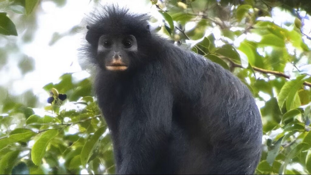
 Colobus congoensis – credit, released by Daniel Rosengren, Frankfurt Zoological Society
When news reaches the public that a new species has been identified, the chances really are 9 times out of 10 that it’s some deep sea slug or a spider.
From the Congo comes the story of the exception—a new species of colobus monkey has been identified, becoming just the 5th such occasion in 75 years of research on the Colorful Continent.
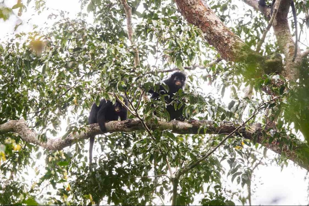
A pair of Colobus congoensis – credit, released by Daniel Rosengren, Frankfurt Zoological Society
The creature with black fur and an innocent face, also bears a striking set of orange lips.
Named Colobus congoensis and known locally as “Likweli” in its home of Lomami National Park, the monkey had lain hidden from our sight within this remote region of the Congo Basin despite decades of scientific exploration in Central Africa.
The mystery of this new species began with an unexpected sighting in 2008, when researchers captured a partially obscured photograph of the monkey. A decade later, researchers encountered the animal again and obtained a much clearer image. That discovery sparked further investigation into the elusive primate.
Now, new genetic, anatomical, and acoustic analyses have confirmed that the monkey represents a distinct evolutionary lineage that diverged from its closest known relative, the black colobus monkey, 4 to 5 million years ago
“This discovery is both exciting and deeply personal, highlighting the extraordinary biodiversity of my homeland and how much remains undocumented,” said Junior Amboko, a Congolese scientist and co-corresponding author of the findings in a statement.
Smaller than related colobus monkeys—about 15 pounds—it is distinguished by sleek, light-reflecting fur and dramatic facial features created by long black facial hairs and large folded ears. White perianal markings further distinguish this species.
In a separate response to the BBC, Amboko said that the animal had a small range compared to other colobus monkeys, suggesting it could be already Endangered. In the statement, it’s detailed that between 2018 and 2022, researchers recorded 114 sightings across an estimated range of around 900 square miles.
“As part of our search, we interviewed people in 52 villages close to where the animals live. And only people in 8 villages [had ever seen] them.”While the scientists’ official recommendation in their paper describing the monkey is that it should be listed as Endangered, the locals also told them that the monkey was a target of local indigenous hunters. New Species of Monkey Found in DR Congo Shows How Much There’s Left to Discover