For many people, the thought of a tasty burger or a cold pint of beer conjures up a vivid mental image and drives behaviour.
This link between thinking and doing serves a clear function – it motivates us to get the necessities for life.
But for some, this process can malfunction. Preoccupation with these rewarding stimuli can lead to disorders of substance overuse, including overeating to the point of obesity and alcohol abuse.
Studies going back to the 1970s have linked vivid mental imagery with drug abuse.
Understanding this link between craving and consuming is central to understanding addiction. This has eluded neuroscience for decades, but the introduction of a new class of drugs for weight loss may have given us just the lever we need to understand it.
These new drugs – including Ozempic and Wegovy – mimic the GLP-1 hormone to stimulate insulin release, slow digestion, and increase feelings of fullness. They are known as GLP-1 agonists and were originally used to treat type 2 diabetes because they help control blood sugar.
As a side effect, people using these drugs also lost a lot of weight, in some cases almost as much as might be expected from bariatric surgery.
But there is another less well publicised effect. Human studies show that GLP-1 agonists reduce alcohol consumption. Preclinical animal studies suggest these drugs also reduce the use of cocaine, amphetamines, opiates and nicotine.
These drugs are changing how we think about the brain’s reward system. They may also open new treatment options for obesity, alcohol dependence and the consumption of other addictive substances.
How the brain regulates reward stimuli
We have a reasonable understanding of the brain’s “reward circuitry” associated with regions that produce the neurotransmitter dopamine.
These brain parts – the ventral tegmental area (VTA) and nucleus accumbens (NAc) – have been the subject of research on reward for decades. They are the obvious candidate regions to look for a mechanism for GLP-1 action in the brain. But they lack significant density of receptors for GLP-1 and are unlikely to be the direct mechanism.
We must, therefore, consider other brain regions to understand the anti-consumption effect of GLP-1 drugs.
One jump “upstream” from the dopamine-producing brain parts is a region called the lateral septum. This brain structure has been historically implicated in emotional regulation.
Back in 1953, pioneering US behavioural researchers Joseph Brady and Walle Nauta coined the term “septal rage” when animals with damage in the lateral septum showed increased aggression, while direct stimulation of this brain region reduced aggression.
Much more recent work has placed the lateral septum at the centre of a neural connectivity network. This has reframed how we think about its function.
While a link between the lateral septum and another region called the hypothalamus is probably responsible for septal rage, the lateral septum links with many other regions with various functions.
The brain’s reward control centre
The lateral septum inherits much of its primary input from a brain region called the hippocampus.
This region is well known as the place that lets us form long-term “episodic memories”. A famous case of hippocampal damage, Henry Molaison (patient HM), was unable to form new memories after his surgery for epilepsy. He effectively lived without a past, in permanent present tense.
The hippocampus also contains the remarkable “place cells” – neurons that fire corresponding to a person’s thoughts about their position in space and, as recent research has shown, time.
This “where and when am I” information gets forwarded to the lateral septum. Key research has recently shown the lateral septum also contains place cells, but these cells strongly respond to rewards. They effectively add “what is good in this place” to the “where and when am I” information from the hippocampus.
Critically, the lateral septum shares this information with the dopamine-producing regions we would normally associate with reward.
Neuroscientists now think of the lateral septum as the brain region that lets us “think about” rewards – our conscious perception of them – and communicates with the machinery in the brain’s reward system that produces dopamine to make us feel good about them.
There is one last reason to suspect the lateral septum as the mechanism behind the anti-consumption effect of GLP-1 agonists. It is absolutely loaded with GLP-1 receptors.
Emerging research points to this as the mechanism. GLP-1 activation directly in the lateral septum has recently been shown to reduce food consumption in mice. Earlier this year, another study showed the same for alcohol consumption.
My own lab has shown this year that GLP-1 drugs reduce a type of activity in the lateral septum that may prevent it communicating so effectively with other brain regions.
These findings are reshaping our understanding of how the brain processes rewards and have put the spotlight firmly on the lateral septum as the home of cravings.![]()
Robert Munn, Senior Lecturer, University of Otago
This article is republished from The Conversation under a Creative Commons license. Read the original article.
 \
\
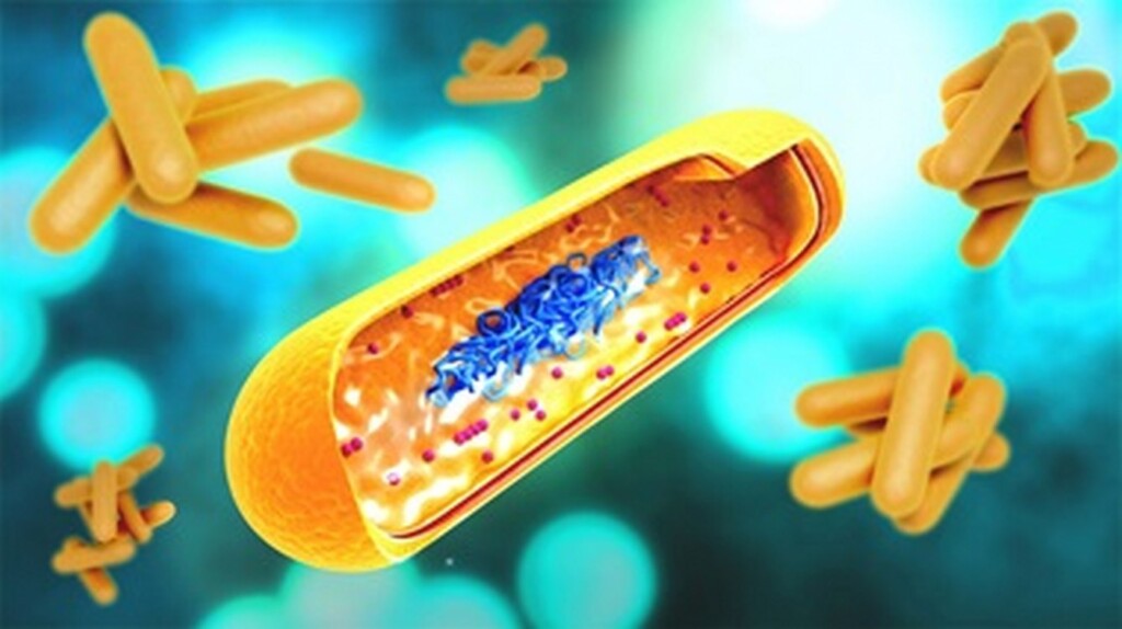
 Macrophages (green) engulfing melanoma cells (purple). Keith et al. / Garvan Institute,


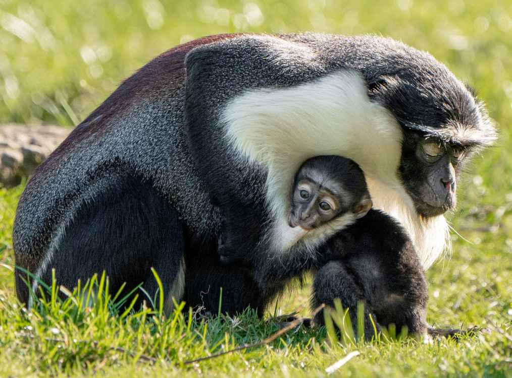

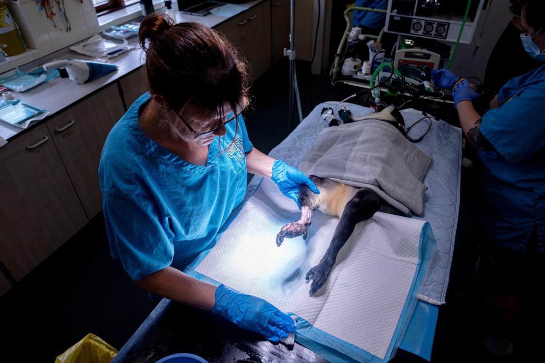 Masaya at the Liverpool Vet. Hospital where she underwent surgery – credit, Chester Zoo via SWNS