The first pig kidney has been transplanted into a living person. But we’re still a long way from solving organ shortages

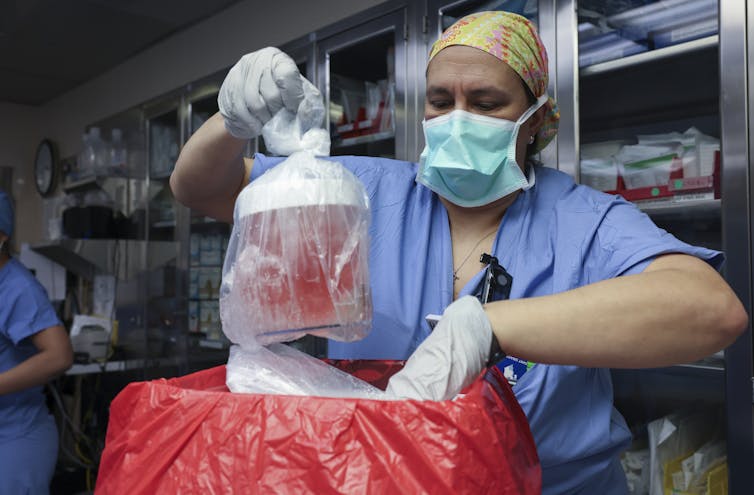
In a world first, we heard last week that US surgeons had transplanted a kidney from a gene-edited pig into a living human. News reports said the procedure was a breakthrough in xenotransplantation – when an organ, cells or tissues are transplanted from one species to another.

The world’s first transplant of a gene-edited pig kidney into a live human was announced last week.

Champions of xenotransplantation regard it as the solution to organ shortages across the world. In December 2023, 1,445 people in Australia were on the waiting list for donor kidneys. In the United States, more than 89,000 are waiting for kidneys.

One biotech CEO says gene-edited pigs promise “an unlimited supply of transplantable organs”.

Not, everyone, though, is convinced transplanting animal organs into humans is really the answer to organ shortages, or even if it’s right to use organs from other animals this way.

There are two critical barriers to the procedure’s success: organ rejection and the transmission of animal viruses to recipients.

But in the past decade, a new platform and technique known as CRISPR/Cas9 – often shortened to CRISPR – has promised to mitigate these issues.

What is CRISPR?

CRISPR gene editing takes advantage of a system already found in nature. CRISPR’s “genetic scissors” evolved in bacteria and other microbes to help them fend off viruses. Their cellular machinery allows them to integrate and ultimately destroy viral DNA by cutting it.

In 2012, two teams of scientists discovered how to harness this bacterial immune system. This is made up of repeating arrays of DNA and associated proteins, known as “Cas” (CRISPR-associated) proteins.

When they used a particular Cas protein (Cas9) with a “guide RNA” made up of a singular molecule, they found they could program the CRISPR/Cas9 complex to break and repair DNA at precise locations as they desired. The system could even “knock in” new genes at the repair site.

In 2020, the two scientists leading these teams were awarded a Nobel prize for their work.

In the case of the latest xenotransplantation, CRISPR technology was used to edit 69 genes in the donor pig to inactivate viral genes, “humanise” the pig with human genes, and knock out harmful pig genes.

How does CRISPR work?

A busy time for gene-edited xenotransplantation

While CRISPR editing has brought new hope to the possibility of xenotransplantation, even recent trials show great caution is still warranted.

In 2022 and 2023, two patients with terminal heart diseases, who were ineligible for traditional heart transplants, were granted regulatory permission to receive a gene-edited pig heart. These pig hearts had ten genome edits to make them more suitable for transplanting into humans. However, both patients died within several weeks of the procedures.

Earlier this month, we heard a team of surgeons in China transplanted a gene-edited pig liver into a clinically dead man (with family consent). The liver functioned well up until the ten-day limit of the trial.

How is this latest example different?

The gene-edited pig kidney was transplanted into a relatively young, living, legally competent and consenting adult.

The total number of gene edits edits made to the donor pig is very high. The researchers report making 69 edits to inactivate viral genes, “humanise” the pig with human genes, and to knockout harmful pig genes.

Clearly, the race to transform these organs into viable products for transplantation is ramping up.

From biotech dream to clinical reality

Only a few months ago, CRISPR gene editing made its debut in mainstream medicine.

In November, drug regulators in the United Kingdom and US approved the world’s first CRISPR-based genome-editing therapy for human use – a treatment for life-threatening forms of sickle-cell disease.

The treatment, known as Casgevy, uses CRISPR/Cas-9 to edit the patient’s own blood (bone-marrow) stem cells. By disrupting the unhealthy gene that gives red blood cells their “sickle” shape, the aim is to produce red blood cells with a healthy spherical shape.

Although the treatment uses the patient’s own cells, the same underlying principle applies to recent clinical xenotransplants: unsuitable cellular materials may be edited to make them therapeutically beneficial in the patient.

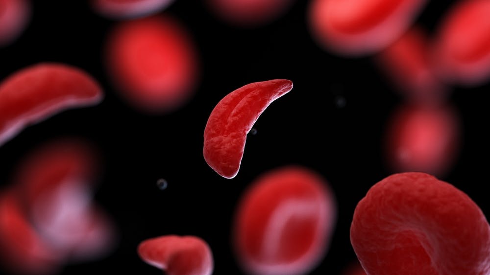
CRISPR technology is aiming to restore diseased red blood cells to their healthy round shape. Sebastian Kaulitzki/Shutterstock

We’ll be talking more about gene-editing

Medicine and gene technology regulators are increasingly asked to approve new experimental trials using gene editing and CRISPR.

However, neither xenotransplantation nor the therapeutic applications of this technology lead to changes to the genome that can be inherited.

For this to occur, CRISPR edits would need to be applied to the cells at the earliest stages of their life, such as to early-stage embryonic cells in vitro (in the lab).

In Australia, intentionally creating heritable alterations to the human genome is a criminal offence carrying 15 years’ imprisonment.

No jurisdiction in the world has laws that expressly permits heritable human genome editing. However, some countries lack specific regulations about the procedure.

Is this the future?

Even without creating inheritable gene changes, however, xenotransplantation using CRISPR is in its infancy.

For all the promise of the headlines, there is not yet one example of a stable xenotransplantation in a living human lasting beyond seven months.

While authorisation for this recent US transplant has been granted under the so-called “compassionate use” exemption, conventional clinical trials of pig-human xenotransplantation have yet to commence.

But the prospect of such trials would likely require significant improvements in current outcomes to gain regulatory approval in the US or elsewhere.

By the same token, regulatory approval of any “off-the-shelf” xenotransplantation organs, including gene-edited kidneys, would seem some way off.The Conversation

Christopher Rudge, Law lecturer, University of Sydney

This article is republished from The Conversation under a Creative Commons license. Read the original article.

Read More........

Scientists Have 3D-Scanned Thousands of Creatures Creating Incredibly Intricate Images Anyone Can Access for Free

3D scanned creatures by oVert – Released by Florida Museum of Natural History / SWNS
An incredible new project has scanned thousands of creatures to advance scientific research and provide colorful images to the world. Natural history museums have entered a new stage of scientific discovery and accessibility with the completion of openVertebrate (oVert), a five-year collaborative project among 18 institutions to create 3D reconstructions of vertebrate specimens and make them freely available online. Now, researchers have published a summary of the project in the journal BioScience reviewing the specimens they’ve scanned to date, offering a glimpse of how the data might be  used to ask newquestions and spur the development of innovative technology. “When people first collected these specimens, they had no idea what the future would hold for them,” said Edward Stanley, co-principal investigator of the oVert project and associate scientist at the Florida Museum of Natural History. Such museums got their start in the 16th century as cabinets of curiosity, in which a few wealthy individuals amassed rare and exotic specimens, which they kept mostly to themselves. Since then, museums have become a resource for the public to learn about biodiversity. But, the majority of museum collections remain behind closed doors—accessible only to scientists who must either travel to see them or ask that a small number of specimens be mailed on loan—and oVert wants to change that. “Now we have scientists, teachers, students and artists around the world using these data remotely,” said David Blackburn, lead principal investigator of the oVert project and curator of herpetology at the Florida Museum. Beginning in 2017, the oVert team members took CT scans of more than 13,000 specimens, with vertebrate species across the tree of life, including over half the genera of all amphibians, reptiles, fishes, and mammals.
A collage of scanned fish from oVert – Released by Florida Museum of Natural History / SWNS
CT scanners use high-energy X-rays to peer past an organism’s exterior and view the dense bone structure beneath. Some specimens were also stained with a contrast-enhancing solution for visualizing soft tissues, like skin, muscle, and other organs. The models give an intimate look at internal portions of a specimen that could previously only be observed through destructive dissection and tissue sampling. “You want to protect specimens, but you also want to have people use them,” Blackburn said. “oVert is a way of reducing the wear and tear on samples while also increasing access, and it’s the next logical step in the
Hedgehog CT scan from oVert – Florida Museum of Natural History / SWNS
mission of museum collections.” Skeletons too large to fit into a CT scanner, like a humpback whale, were painstakingly taken apart so that 3D models of each individual bone could be scanned and reassembled. “These are not things you put in boxes and loan,” Blackburn pointed out. A set of iconic Galapagos tortoises at the California Academy of Sciences were each photographed in a 360-degree rotation. Photographing their undersides was problematic, as their curved shells made it impossible to keep them upright. After a few trial-and-error runs, they settled on placing the specimens on top of inflatable swimming tubes. Scientists have already used data from the project to gain astonishing insights into the natural world. Watch the incredible video below, and learn more at the bottom…In 2023, Edward Stanley was conducting routine CT scans of spiny mice and was surprised to find their tails were covered with an internal coat of bony plates, called osteoderms. Before this discovery, armadillos were considered to be the only living mammals with these structures. “All kinds of things jump out at you when you’re when you’re scanning,” Stanley said. “I study osteoderms, and through kismet or fate, I happened to be the one scanning those particular specimens on that particular day and noticed something strange about their tails on the X-ray. “That happens all the time. We’ve found all sorts of strange, unexpected things.”oVert scans were used to determine what killed a rim rock crown snake, considered to be the rarest snake species in North America. Another study showed that a group of frogs called pumpkin toadlets had become so small that the fluid-filled canals in their ears that confer balance no longer functioned properly, causing them to crash-land when jumping. One study of 500 oVert specimens revealed that frogs have lost and regained teeth more than 20 times throughout their evolutionary history. Other researchers concluded that Spinosaurus, a massive dinosaur that was larger than Tyrannosaurus rex and thought to be aquatic, would have actually been a poor swimmer, and thus likely stayed on land. And the list goes on, full of insights and ideas that would have been impossible or impractical before the project’s outset. “Now that we’ve been working on this for so long, we have a broad scaffold that allows us to take a broader view of
Fish CT scan from oVert – Florida Museum of Natural History / SWNS
evolutionary questions,” Stanley said. Artists and teachers are benefitting too Funded in part by the National Science Foundation, the value of the oVert project extends beyond science. Artists have used the 3D models to create realistic animal replicas, photographs of oVert specimens have been displayed as museum exhibits, and specimens have been incorporated into virtual reality headsets that give users the chance to interact with and manipulate them. A high school teacher in Cincinnati says it’s been a game-changer for her studies on evolution. “I teach juniors and seniors, and I absolutely love them, but they can be a tough audience,” said Jennifer Broo. “They know when things are fake, which makes them less engaged. Using the oVert models, my class has gotten so much better because I have had the opportunities to work with and expose my students to real data.”Visit Sketchfab to view a sample of 3D interactive models. At MorphoSource you can access the full openVertebrate repository.Scientists Have 3D-Scanned Thousands of Creatures Creating Incredibly Intricate Images Anyone Can Access for Free
Read More........