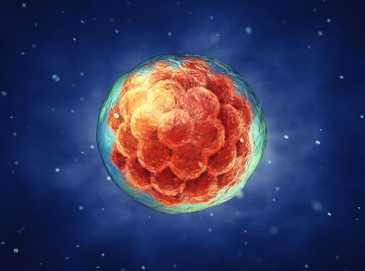
Pigs with human brain cells and biological chips: how lab-grown hybrid lifeforms bamboozle scientific ethics


Earlier this month, scientists at the Guangzhou Institutes of Biomedicine and Health announced they had successfully grown “humanised” kidneys inside pig embryos.

The scientists genetically altered the embryos to remove their ability to grow a kidney, then injected them with human stem cells. The embryos were then implanted into a sow and allowed to develop for up to 28 days.

The resulting embryos were made up mostly of pig cells (although some human cells were found throughout their bodies, including in the brain). However, the embryonic kidneys were largely human.

This breakthrough suggests it may soon be possible to generate human organs inside part-human “chimeric” animals. Such animals could be used for medical research or to grow organs for transplant, which could save many human lives.

But the research is ethically fraught. We might want to do things to these creatures we would never do to a human, like kill them for body parts. The problem is, these chimeric pigs aren’t just pigs – they are also partly human.

If a human–pig chimera were brought to term, should we treat it like a pig, like a human, or like something else altogether?

Maybe this question seems too easy. But what about the idea of creating monkeys with humanised brains?

Chimeras are only one challenge among many

Other areas of stem cell science raise similarly difficult questions.

In June, scientists created “synthetic embryos” – lab-grown embryo models that closely resemble normal human embryos. Despite the similarities, they fell outside the scope of legal definitions of a human embryo in the United Kingdom (where the study took place).

Like human–pig chimeras, synthetic embryos straddle two distinct categories: in this case, stem cell model and human embryo. It is not obvious how they should be treated.

In the past decade, we have also seen the development of increasingly sophisticated human cerebral organoids (or “lab-grown mini-brains”).

Unlike synthetic embryos, cerebral organoids don’t mimic the development of a whole person. But they do mimic the development of the part that stores our memories, thinks our thoughts, and makes conscious experience possible.

A network of neural cells grown on an array of electrodes to produce a ‘biological computer chip’. Cortical Labs

Most scientists think current “mini-brains” are not conscious, but the field is developing rapidly. It is not far-fetched to think a cerebral organoid will one day “wake up”.

Complicating the picture even further are entities that combine human neurons with technology – like DishBrain, a biological computer chip made by Cortical Labs in Melbourne.

How should we treat these in vitro brains? Like any other human tissue culture, or like a human person? Or perhaps something in between, like a research animal?

A new moral framework

It might be tempting to think we should settle these questions by slotting these entities into one category or another: human or animal, embryo or model, human person or mere human tissue.

This approach would be a mistake. The confusion sparked by chimeras, embryo models, and in vitro brains shows these underlying categories no longer make sense.

We are creating entities that are neither one thing nor the other. We cannot solve the problem by pretending otherwise.

We would also need good reasons to classify an entity one way or another.

Should we count the proportion of human cells to determine whether a chimera counts as an animal or a human? Or should it matter where the cells are located? What matters more, brain or buttocks? And how can we work this out?

Moral status

Philosophers would say these are questions about “moral status”, and they have spent decades deliberating on what kinds of creatures we have moral duties to, and how strong these duties are. Their work can help us here.

For example, utilitarian philosophers see moral status as a matter of whether a creature has any interests (in which case it has moral status), and how strong those interests are (stronger interests matter more than weaker ones).

On this view, so long as an embryo model or brain organoid lacks consciousness, it will lack moral status. But if it develops interests, we need to take these into account.

Similarly, if a chimeric animal develops new cognitive abilities, we need to reconsider our treatment of it. If a neurological chimera comes to care about its life as much as a typical human does, then we should hesitate to kill it just as much as we would hesitate to kill a human.

This is just the beginning of a bigger discussion. There are other accounts of moral status, and other ways of applying them to the entities stem cell scientists are creating.

But thinking about moral status sets us down the right path. It fixes our minds on what is ethically significant, and can begin a conversation we badly need to have.The Conversation

Julian Koplin, Lecturer in Bioethics, Monash University & Honorary fellow, Melbourne Law School, Monash University

This article is republished from The Conversation under a Creative Commons license. Read the original article.

Read More........

How consciousness may rely on brain cells acting collectively – new psychedelics research on rats

Psychedelics can help uncover consciousness. agsandrew/Shutterstock Pär Halje, Lund University
Psychedelics are known for inducing altered states of consciousness in humans by fundamentally changing our normal pattern of sensory perception, thought and emotion. Research into the therapeutic potential of psychedelics has increased significantly in the last decade. While this research is important, I have always been more intrigued by the idea that psychedelics can be used as a tool to study the neural basis of human consciousness in laboratory animals. We ultimately share the same basic neural hardware with other mammals, and possibly some basic aspects of consciousness, too. So by examining what happens in the brain when there’s a psychedelically induced change in conscious experience, we can perhaps glean insights into what consciousness is in the first place.We still don’t know a lot about how the networks of cells in the brain enable conscious experience. The dominating view is that consciousness somehow emerges as a collective phenomenon when the dispersed information processing of individual neurons (brain cells) is integrated as the cells interact.But the mechanism by which this is supposed to happen remains unclear. Now our study on rats, published in Communications Biology, suggests that psychedelics radically change the way that neurons interact and behave collectively.Our study compared two different classes of psychedelics in rats: the classic LSD type and the less-typical ketamine type (ketamine is an anaesthetic in larger doses). Both classes are known to induce psychedelic experiences in humans, despite acting on different receptors in the brain. Exploring brain waves: We used electrodes to simultaneously measure electrical activity from 128 separate areas of the brain of nine awake rats while they were given psychedelics. The electrodes could pick up two kinds of signals: electrical brain waves caused by the cumulative activity in thousands of neurons, and smaller transient electrical pulses, called action potentials, from individual neurons. The classic psychedelics, such as LSD and psilocybin (the active ingredient in magic mushrooms), activates a receptor in the brain (5-HT2A) which normally binds to serotonin, a neurotransmitter that regulates mood and many other things. Ketamine, on the other hand, works by inhibiting another receptor (NMDA), which normally is activated by glutamate, the primary neurotransmitter in the brain for making neurons fire. We speculated that, despite these differences, the two classes of psychedelics might have similar effects on the activity of brain cells. Indeed, it turned out that both drug classes induced a very similar and distinctive pattern of brain waves in multiple brain regions. The brain waves were unusually fast, oscillating about 150 times per second. They were also surprisingly synchronised between different brain regions. Short bursts of oscillations at a similar frequency are known to occur occasionally under normal conditions in some brain
Brain waves on electroencephalogram EEG. Chaikom/Shutterstock
regions. But in this case, it occurred for prolonged durations.  First, we assumed that a single brain structure was generating the wave and that it then spread to other locations. But the data was not consistent with that scenario. Instead, we saw that the waves went up and down almost simultaneously in all parts of the brain where we could detect them – a phenomenon called phase synchronisation. Such tight phase synchronisation over such long distances has to our knowledge never been observed before. We were also able to measure action potentials from individual neurons during the psychedelic state. Action potentials are electrical pulses, no longer than a thousandth of a second, that are generated by the opening and closing of ion channels in the cell membrane. The action potentials are the primary way that neurons influence each other. Consequently, they are considered to be the main carrier of information in the brain. However, the action potential activity caused by LSD and ketamine differed significantly. As such, they could not be directly linked to the general psychedelic state. For LSD, neurons were inhibited – meaning they fired fewer action potentials – in all parts of the brain. For ketamine, the effect depended on cell type – certain large neurons were inhibited, while a type of smaller, locally connecting neurons, fired more. Therefore, it is probably the synchronised wave phenomenon – how the neurons behave collectively – that is most strongly linked to the psychedelic state. Mechanistically, this makes some sense. It is likely that this type of increased synchrony has large effects on the integration of information across neural systems that normal perception and cognition rely on. I think that this possible link between neuron-level system dynamics and consciousness is fascinating. It suggests that consciousness relies on a coupled collective state rather than the activity of individual neurons – it is greater than the sum of its parts. That said, this link is still highly speculative at this point. That’s because the phenomenon has not yet been observed in human brains. Also, one should be cautious when extrapolating human experiences to other animals – it is of course impossible to know exactly what aspects of a trip we share with our rodent relatives. But when it comes to cracking the deep mystery of consciousness, every bit of information is valuable. Pär Halje, Associate Research Fellow of Neurophysiology, Lund University This article is republished from The Conversation under a Creative Commons license. Read the original article.
Read More........

How grandmothers' brains react to the sight of their grandchildren

"We're highlighting the brain functions of grandmothers that may play an important role in our social lives and development," says Minwoo Lee, an Emory graduate student and co-author of the study. "It's an important aspect of the human experience that's been largely left out of the field of neuroscience."

By Carol Clark: Many people lucky enough to have grown up with doting grandmothers know that they can burnish a child’s development in unique and valuable ways. Now, for the first time, scientists have scanned grandmothers’ brains while they’re viewing photos of their young grandchildren — providing a neural snapshot of this special, inter-generational bond.

Proceedings of the Royal Society B published the first study to examine grandmaternal brain function, conducted by researchers at Emory University.

“What really jumps out in the data is the activation in areas of the brain associated with emotional empathy,” says James Rilling, lead author and professor in Emory's Department of Anthropology and Department of Psychiatry and Behavioral Sciences. “That suggests that grandmothers are geared toward feeling what their grandchildren are feeling when they interact with them. If their grandchild is smiling, they’re feeling the child’s joy. And if their grandchild is crying, they’re feeling the child’s pain and distress.”

In contrast, the study found that when grandmothers view images of their adult child, they show stronger activation in an area of the brain associated with cognitive empathy. That indicates they may be trying to cognitively understand what their adult child is thinking or feeling and why, but not as much from the emotional side.

“Young children have likely evolved traits to be able to manipulate not just the maternal brain, but the grand maternal brain,” Rilling says. “An adult child doesn’t have the same cute ‘factor,’ so they may not illicit the same emotional response.”

Co-authors of the study are Minwoo Lee, a PhD candidate in Emory’s Department of Anthropology, and Amber Gonzalez, a former Emory research specialist.
"What really jumps out in the data is the activation in areas of the brain associated with emotional empathy," Rilling says.

“I can relate to this research personally because I spent a lot of time interacting with both of my grandmothers,” Lee says. “I still remember warmly the moments I had with them. They were always so welcoming and happy to see me. As a child, I didn’t really understand why.”

It’s relatively rare, Lee adds, for scientists to study the older human brain outside of the problems of dementia or other aging disorders.

“Here, we’re highlighting the brain functions of grandmothers that may play an important role in our social lives and development,” Lee says. “It’s an important aspect of the human experience that has been largely left out of the field of neuroscience.”

Rilling’s lab focuses on the neural basis of human social cognition and behavior. Motherhood has been extensively studied by other neuroscientists. Rilling is a leader in researching the lesser-explored neuroscience of fatherhood.

Grandmothers interacting with grandchildren offered new neural territory.

“Evidence is emerging in neuroscience for a global, parental caregiving system in the brain,” Rilling says. “We wanted to see how grandmothers might fit into that pattern.”

Humans are cooperative breeders, meaning that mothers get help caring for their offspring, although the sources of that help vary both across and within societies.

“We often assume that fathers are the most important caregivers next to mothers, but that’s not always true,” Rilling says. “In some cases, grandmothers are the primary helper.”

In fact, the “grandmother hypothesis” posits that the reason human females tend to live long past their reproductive years is because they provide evolutionary benefits to their offspring and grandchildren. Evidence supporting this hypothesis includes a study of the traditional Hadza people of Tanzania, where foraging by grandmothers improves the nutritional status of their grandchildren. Another study of traditional communities showed that the presence of grandmothers decreases their daughters’ interbirth intervals and increases the number of grandchildren.

And in more modern societies, evidence is accumulating that positively engaged grandmothers are associated with children having better outcomes on a range of measures, including academic, social, behavior and physical health.
"If their grandchild is smiling, they're feeling the child's joy," Rilling says. "And if their grandchild is crying, they're feeling the child's pain and distress."

For the current study, the researchers wanted to understand the brains of healthy grandmothers and how that may relate to the benefits they provide to their families.

The 50 participants in the study completed questionnaires about their experiences as grandmothers, providing details such as how much time they spend with their grandchildren, the activities they do together and how much affection they feel for them. They also underwent functional magnetic resonance imaging (fMRI) to measure their brain function as they viewed pictures of their grandchild, an unknown child, the same-sex parent of the grandchild, and an unknown adult.

The results showed that, while viewing pictures of their grandchildren, most participants showed more activity in brain areas involved with emotional empathy and movement, compared to when they were viewing the other images.

Grandmothers who more strongly activated areas involved with cognitive empathy when viewing pictures of their grandchild reported in the questionnaire that they desired greater involvement in caring for the grandchild.

Finally, compared with results from earlier study by the Rilling lab of fathers viewing photos of their children, grandmothers more strongly activated regions involved with emotional empathy and motivation, on average, when viewing images of their grandchildren.

“Our results add to the evidence that there does seem to be a global parenting caregiving system in the brain, and that grandmothers’ responses to their grandchildren maps onto it,” Rilling says.

One limitation to the study, the researchers note, is that the participants skewed towards mentally and physically healthy women who are high-functioning grandmothers.

The study opens the door to many more questions to be explored. “It would be interesting to also look at the neuroscience of grandfathers and how the brain functions of grandparents may differ across cultures,” Lee says.

An especially gratifying aspect of the project for Rilling was personally interviewing all the participants himself. “It was fun,” he says. “I wanted to get a sense of the rewards and challenges of being a grandmother.”

The main challenge many of them reported was trying not to interfere when they disagreed with the parents over how their grandchildren should be raised and what values should be instilled in them.

“Many of them also said how nice it is to not be under as much time and financial pressure as they were when raising their children,” Rilling says. “They get to enjoy the experience of being a grandmother much more than they did being parents.” This work was supported in part by the Silvia O. Conte Center for Oxytocin and Social CognitionSource eScienceCommons:
Read More........